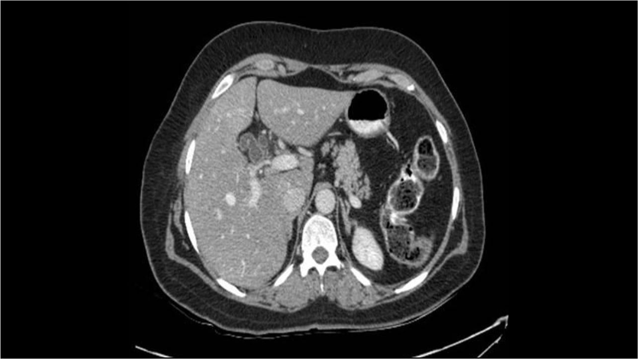
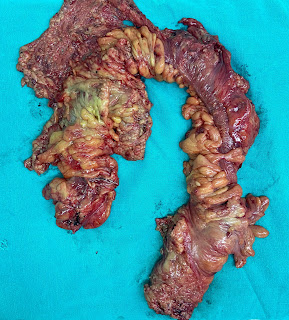
A nonsmoker, non alcoholic elderly woman presented to the outpatients’ clinic with 2 weeks history of postprandial pain radiating to back. She denied vomiting or weight loss. Her general physical and abdominal examinations were unremarkable.
The initial hematologic, biochemical and liver function tests were within prescribed normal range.
Following ultrasound examination, abdominal contrast enhanced computed tomography (CECT) scan was done (Figure 1 & 2) which shows a normally enhancing liver with no space occupying lesion or intrahepatic biliary radical dilatation. There is a cystic dilatation of common duct. The gallbladder shows an enhancing polypoidal mass lesion. The fat planes between the liver and gallbladder are well preserved. The pancreas and spleen appear normal and there is no significant lymphadenopathy.
In view of abdominal CECT findings, a diagnosis of choledochal cyst type I (cystic) with probable malignant gallbladder mass lesion was made.
A radical cholecystectomy with resection of common duct was performed. The histopathology was suggestive of choledochal cyst with papillary carcinoma (T1bN0M0) of the gallbladder. In adult patients with choledochal cyst, the incidence of carcinoma is reported to be 11.4% with median age of diagnosis being 42 years. Cholangiocarcinoma (70.4%) followed by gallbladder cancer (23.5%) are the most common malignancies.


